Causes of Low or High White Blood Cells Biology Diagrams This human anatomy diagram with labels depicts and explains the details and or parts of the Human Spleen.Human anatomy diagrams and charts show internal organs, body systems, cells, conditions, sickness and symptoms information and/or tips to ensure one lives in good health. This human anatomy diagram with labels depicts and explains the details and or parts of the Spleen In Humans. Human anatomy diagrams and charts show internal organs, body systems, cells, conditions, sickness and symptoms information and/or tips to ensure one lives in good health. One of the spleen's main jobs is to filter your blood. To best depict the location of the spleen, we'll describe its relations. The spleen is found in the left hypochondriac region of the abdomen (left upper quadrant). More precisely, the spleen is located posterior to the stomach and anterior to the left hemidiaphragm at the level of ribs 9-10. Medial to the spleen is the left kidney; superior is the diaphragm, while inferiorly it rests
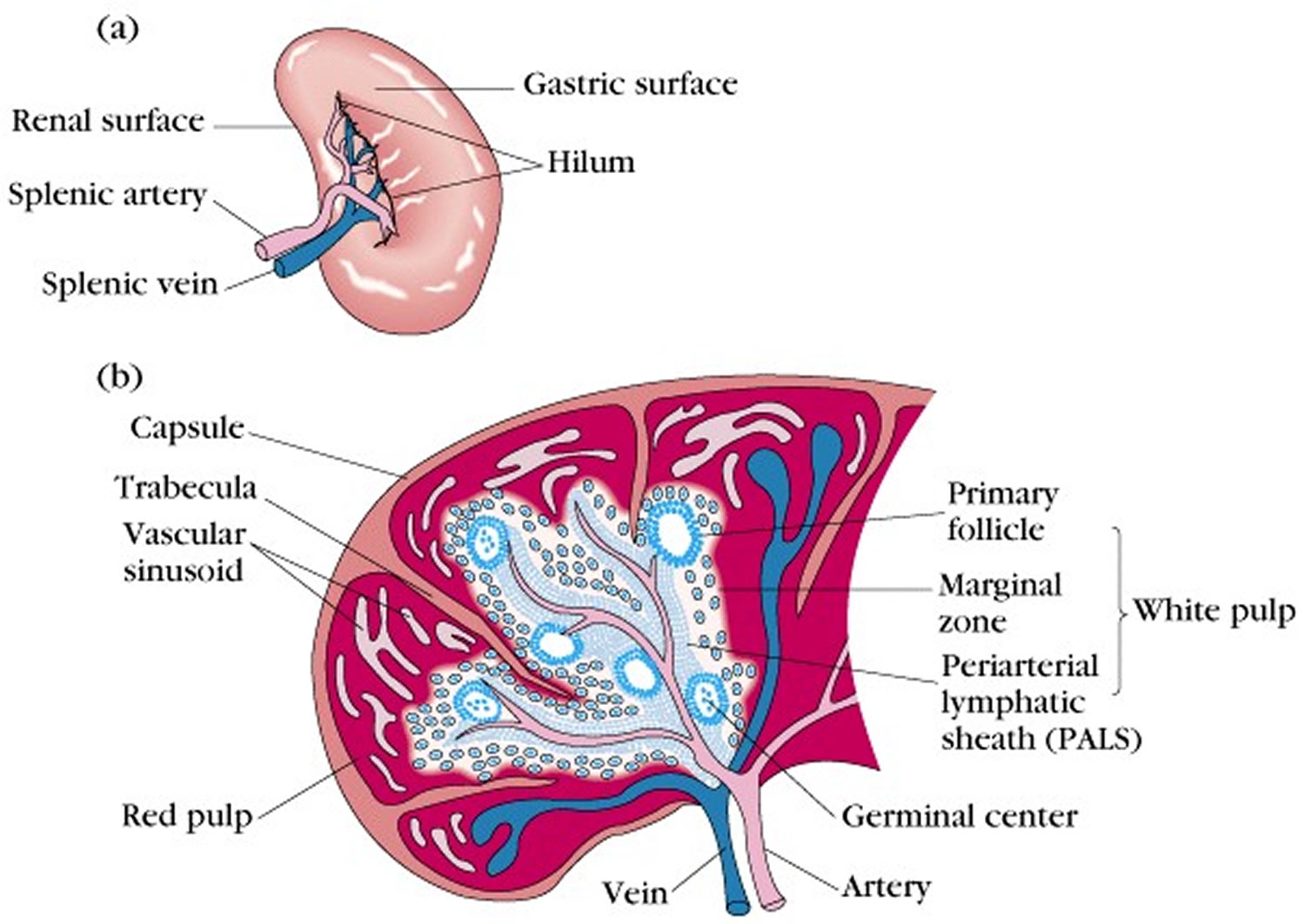
Spleen histology slide (labeled) The spleen is a fist sized organ located in the left upper quadrant of the abdomen. It is the largest lymphoid organ and thus the largest filter of blood in the human body. The spleen has a unique location, embryological development and histological structure that differs significantly from other lymphoid organs. Gross and Microscopic Anatomy . The spleen is a reticuloendothelial lymphoid organ located in the left upper abdomen, posterolateral to the stomach, tail of the pancreas, and colic flexure. The adult spleen is 10 to 12 cm in length and weighs an average of 168 g in the adult man and 135 g in the adult woman. The diagram depicts the Structure and Anatomy. The spleen is a soft, highly vascular, fist-sized organ that plays a vital role in the lymphatic and circulatory systems. It is encased in a fibrous capsule and consists of two main types of tissue—red pulp and white pulp—which perform distinct functions related to blood filtration and immune response. Below is a
3D Interactive Anatomy Tutorials Biology Diagrams
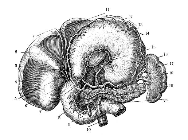
The spleen is a small organ inside your left rib cage, just above the stomach. It's part of the lymphatic system (which is part of the immune system). The spleen stores and filters blood and makes white blood cells that protect you from infection. Many diseases and conditions can affect how the spleen works. A ruptured (torn) spleen can be fatal. The spleen is an organ located in the upper left abdomen, roughly the size of a clenched fist. In the adult, the spleen functions mainly as a blood filter, removing old red blood cells. In this article, we shall look at the anatomy of the spleen - its anatomical position, structure and vasculature. By TeachMeSeries Ltd (2025) Fig 1 The
Learn about the anatomy of the spleen including its anatomical relations and vascular supply using these interactive modules and video tutorials ADVERTISEMENTS: In this article we will discuss about the structure of spleen with the help of suitable diagram. Also learn about its functions. Spleen (lien) is the largest lymphoid tissue in the body and specialised, been-shaped organ for filtering blood. It is a highly vascular haemopoietic organ situated in the left hypochondrium directly beneath the […]