fascia anatomy trains Google Search Biology Diagrams Fascia is a connective tissue that supports, surrounds, and provides shape for nerves, muscles, tendons, and joints. This type of collagen comprises cell membranes, hair, and the human placenta. All the different types of collagen are intertwined, providing support to the structures within your body. Quantitative anatomy, radiographic Fascia: A layer of connective tissue that plays an active role in the body. It supports tissues and organs, lessens friction, or eases muscle tension. Learn more in this guide.

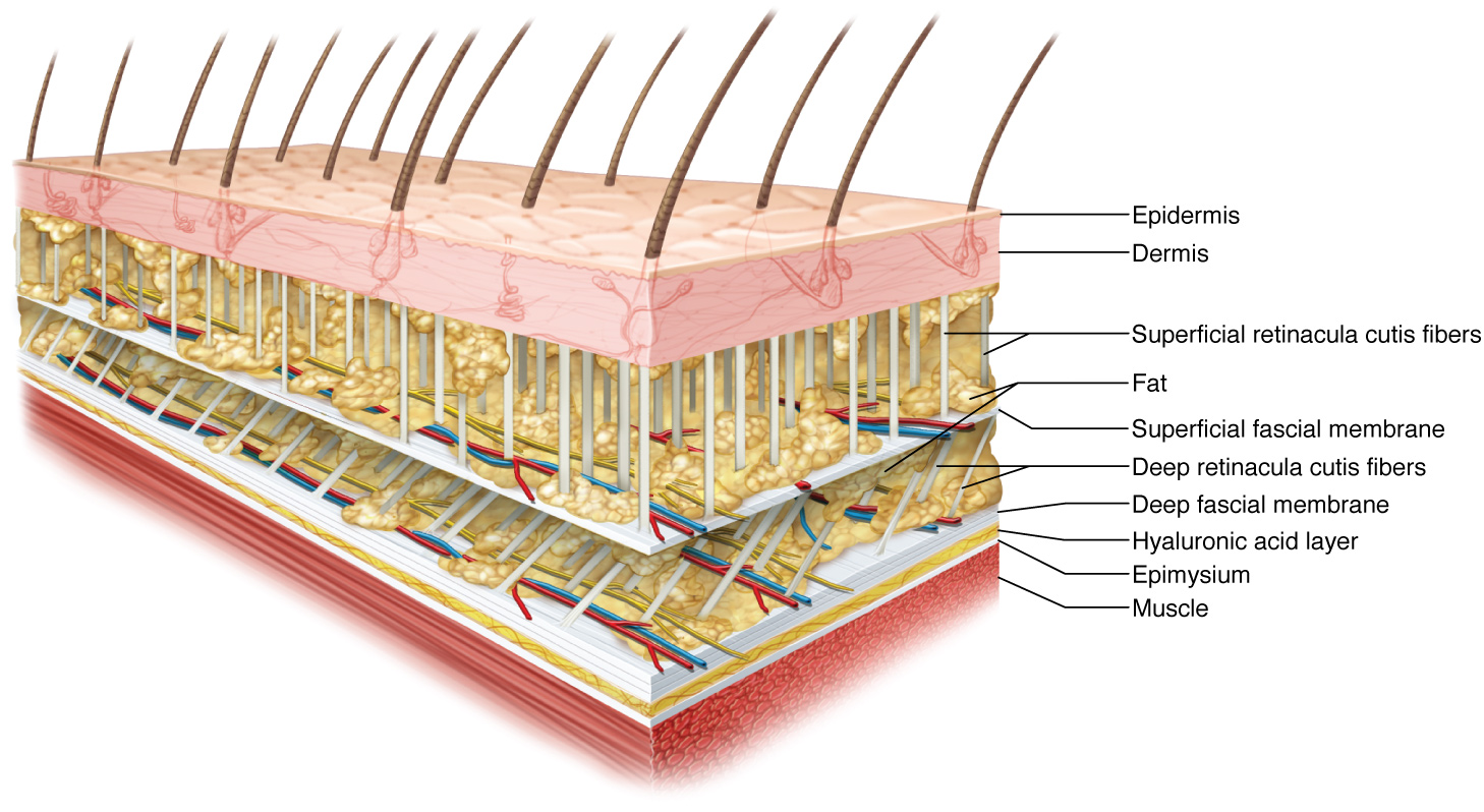
In general, there are two types of fascia: Superficial fascia; Deep fascia; The superficial fascia (i.e. tela subcutanea, hypodermis, subcutaneous tissue) is used to describe the connective that separates the skin from the underlying muscle tissue. The deep fascia is a dense, organized, connective tissue located deep to the skin and Scientists initially thought fascia only provided support to your organs, muscles and bones. Recently, the definition has expanded to include the tissue that surrounds all of the cells, nerves, joints, tissues, tendons and ligaments throughout your body as well. As part of this discovery, they learned that fascia is a part of a system-wide network that provides form and function to every part Researchers do not agree on one comprehensive "fascia" definition. Despite the scientific uncertainty, there is an agreement with medical text that the fascia covers every structure of the body, creating a structural continuity that gives form and function to every tissue and organ. The fascial tissue has a ubiquitous distribution in the body system; it is able to wrap, interpenetrate, support
Fascia Anatomy & Physiology Biology Diagrams
A fascial compartment is a section within the body that contains muscles and nerves and is surrounded by fascia. In the human body, the limbs can each be divided into two segments: Educating Interconnected Anatomy). [8] This plastinate provides a detailed view of the human fascial network, allowing for a better understanding of its The deep fascia, according to this definition, lies below the superficial fascia, highlighting two fasciae. In 2011, the Federative International Programme on Anatomical Terminologies (FIPAT), in agreement with FCAT, defined the fascia as "a sheath, a sheet, or any other dissectible aggregations of connective tissue that forms beneath the

Fascia is made up primarily of collagen, an abundant protein that constitutes about one-third of all protein in the human body and that accounts for most of the content of tendons and ligaments. Hyaluronan ( hyaluronic acid ), a lubricating polysaccharide occurring in the extracellular matrix, lies between each layer of fascia. Fascia is the tensional, continuous fibrillar network within the body, extending from the surface of the skin to the nucleus of the cell. This global network is mobile, adaptable, fractal, and irregular. It constitutes the basic structural architecture of the human body. Fascia is made up of sheets of connective tissue that is found below the skin. These tissues attach, stabilize, impart strength, maintain vessel patency, separate muscles, and enclose different organs. Traditionally, the word fascia was used primarily by surgeons to describe the dissectible tissue seen in the body encasing other organs, muscles, and bones. Recently, the definition has been