Image Of Osteoporosis Stock Photos Image Of Osteoporosis Stock Images Biology Diagrams Human infants typically have 270 bones, fusing into around 206 in the human adult. Variability in number arises from some bones' anatomic variations. Bones differ in size, shape, and strength, depending on function. [2] Understanding bone anatomy and physiology helps healthcare professionals treat skeletal conditions. Explore the skeletal system with our interactive 3D anatomy models. Learn about the bones, joints, and skeletal anatomy of the human body.
At the end of this unit, you should be able to: I. Describe the functions of the skeletal system and the five basic shapes of human bones. II. Describe the structure and histology of the skeletal system. III. Define and identify the following parts of a long bone: diaphysis, epiphysis, metaphysis, articular cartilage, periosteum, medullary cavity, and endosteum. IV. Compare the composition and 3D Anatomy Atlas. Explore Human Body in Real Time. By Dr. R. Blanco Salado Download Homepage
TRU Human Anatomy & Physiology I Biology Diagrams
There are 12 major anatomy systems: Skeletal, Muscular, Cardiovascular, Digestive, Endocrine, Nervous, Respiratory, Immune/Lymphatic, Urinary, Female Reproductive

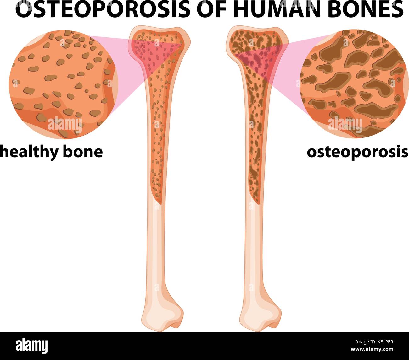
Osteoporosis (OP), which is a common skeletal disease with different causes, is prevalent in the aging population. Postmenopause women generally suffer from OP with bone loss due to estrogen deficiency. Diabetes is also associated with OP by complex metabolic mechanisms. Bone qualities of OP caused by aging were compared with those of the ovariectomy (OVX) model and the Type 2 diabetic model
Osteoporosis : Anatomy & Physiology Biology Diagrams
The BioDigital Human is the first cloud based virtual model of the human body - 3D human anatomy, disease and treatment, all in interactive 3D.
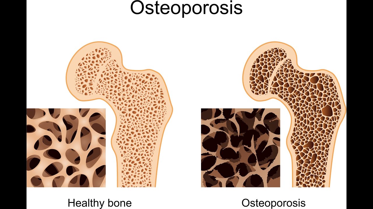
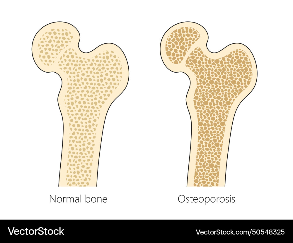
Osteoporosis Osteoporosis refers to a group of diseases in which bone resorption outpaces bone deposit (i.e. bone loss occurs faster than bone growth). The bones become so fragile that something as simple as a hefty sneeze or stepping off a curb can cause them to break. The human skeleton of an adult usually consists of around 206 bones, depending on the counting of Sternum (which may alternatively be included as the manubrium, body of sternum, and the xiphoid process). [1] It is composed of 270 bones at the time of birth, [2] but later decreases to 206: 80 bones in the axial skeleton and 126 bones in the appendicular skeleton. 172 of 206 bones are part of a

List of bones of the human skeleton Biology Diagrams
This article covers the anatomy of all types of human bones, their functions and clinical aspects. Learn about this topic at Kenhub!