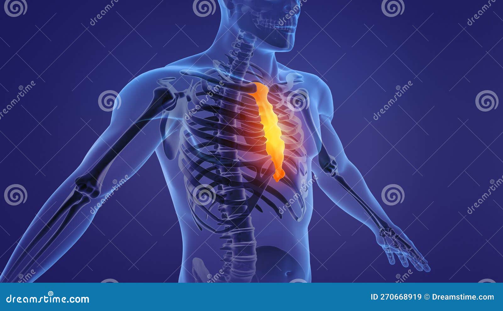
SOLUTION Detailed ppt of Ribs Sternum Biology Diagrams The sternum, commonly referred to as the breastbone, is a flat, elongated bone located in the central part of the chest. It forms the anterior portion of the thoracic cage and serves as an attachment site for the ribs and clavicles.[1] The sternum consists of three parts: the manubrium, body (or gladiolus), and xiphoid process. Through cartilage, it joins other bones and muscles. Your heart and lungs are protected by this part of your rib cage. Your sternum and breastbone are interchangeable. Anatomy of the Sternum. The breastbones are situated in the middle and front of your chest. Sternum appears in the upper region of your chest, exactly on the front of your thymus. False ribs are the next 3 pairs (8th, 9th, and 10th ribs). Their costal cartilages join with that of the 7th rib to indirectly connect to the sternum. Floating ribs are the last 2 pairs (11th and 12th ribs) with no articulation with the sternum. The second mode of classifying ribs is based on their structure and anatomy variations.

What are the parts of the sternum? Your sternum anatomy consists of three bony parts. These parts include: Costochondritis is a condition that causes inflammation in the cartilage that attaches your ribs to your sternum. An infection, injury or arthritis can cause the condition. Costochondritis causes sharp, stabbing rib pain and tenderness.

Anatomy, Location, & Labeled Diagram Biology Diagrams
The sternum is the bone that lies in the anterior midline of our thorax. It forms part of the rib cage and the anterior-most part of the thorax. Its functions are to protect the thoracic organs from trauma and also form the bony attachment for various muscles. It is also the center around which the superior 10 ribs directly or indirectly attached.
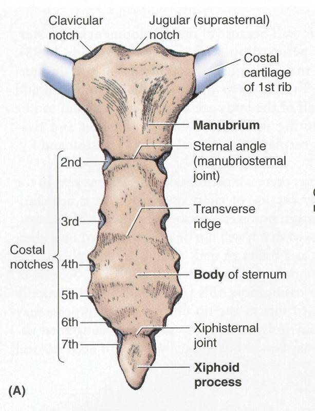
The Sternum Explore the anatomy, structure, and role of the sternum with Innerbody's interactive 3D model. by Tim Taylor Last At its inferior end, the manubrium meets the body of the sternum at the joint with the costal cartilage of the second ribs. Here it forms the sternal angle, a slight posterior bend in the sternum that can be felt

Rib Cage Anatomy: Complete Guide with Parts, Names & Diagram Biology Diagrams
The sternum is a partially T-shaped vertical bone that forms the anterior portion of the chest wall centrally. The sternum is divided anatomically into three segments: manubrium, body, and xiphoid process. The sternum connects the ribs via the costal cartilages forming the anterior rib cage. The manubrium is the broad superior segment, the body is the middle portion, and the xiphoid process is

Overview of Rib Cage Anatomy. The rib cage is an essential body element that protects the heart and lungs. It creates the chest area known as the thorax, which comprises ribs, the sternum, and a portion of the spine. The rib cage anatomy has 24 ribs, 12 on each side, as well as 12 vertebrae, which are bones that form the spine in the chest area.

Structure, Location, Anatomy, Function, Diagram Biology Diagrams
The thoracic cage is a bony case consisting of ribs and sternum which encases vital organs like the lungs and the heart and shapes the chest. There are 12 pairs of ribs in the body. They are attached to the vertebral column with most of the ribs anteriorly joining to the sternum bone via costal cartilage. The first seven ribs directly attach to Parts and Anatomy. As stated, the sternum is a long, flat bone divided into three parts. Its shape can be compared to an upside-down sword. Its top rectangular part, (i) the manubrium, resembles the handle, whereas the flat, long middle portion or (ii) body looks like the sword's blade. The joint between the 1 st rib and the sternum is