The Anatomical Structure Of Skin Layer Biology Diagrams The layers of the skin make up the body's largest organ, providing a crucial barrier between the internal structures and the external environment.This complex, multi-layered tissue is essential for protection, sensation, temperature regulation, and immune defense. Understanding the structure and function of the layers of the skin is key to appreciating its role in human health and physiology.

This article will discuss the anatomy of the skin, including its structure, function, embryology, blood, lymphatic, and nerve supply, surgical, and clinical significance.[1][2] The multiple layers of the skin are dynamic, shedding and replacing old inner layers. The thickness of skin varies based on its location, age, gender, medications

Functions of the Skin Biology Diagrams
Learn about the function, gross structure and ultrastructure of the skin, the largest organ in the human body. Explore the layers of the epidermis, dermis and hypodermis, and the skin appendages and disorders.
Learn about the structure and functions of the epidermis, dermis, and hypodermis, the three layers of the skin. See diagrams, micrographs, and animations of the skin cells and tissues.
Anatomy, Skin (Integument), Epidermis Biology Diagrams
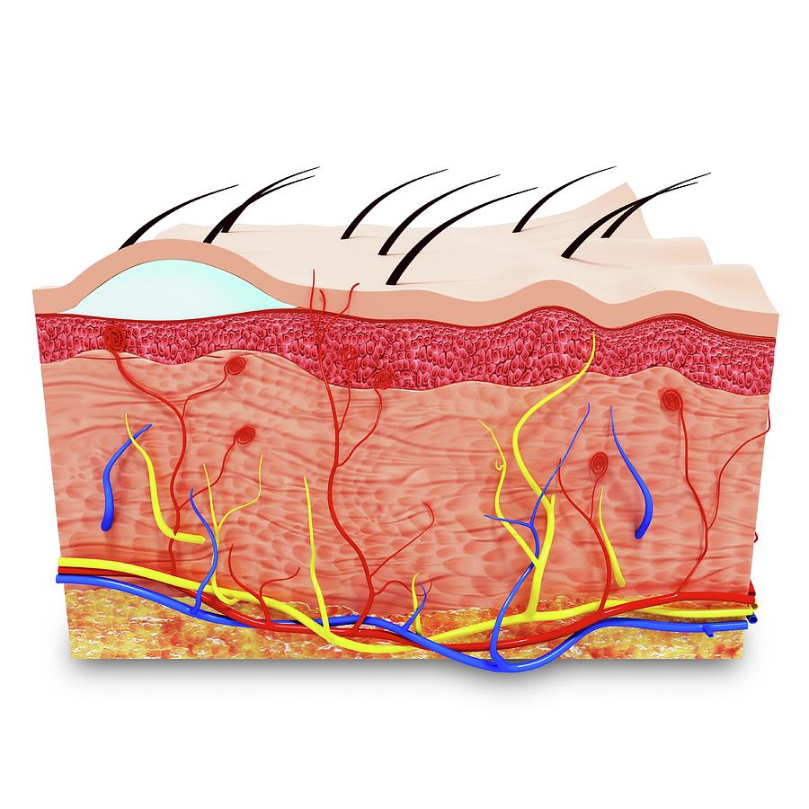
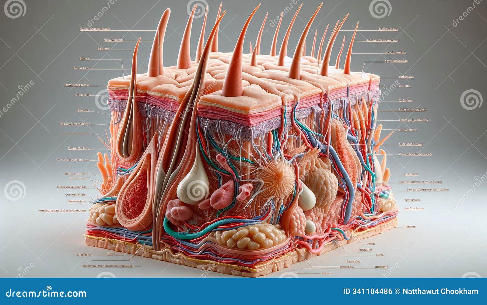
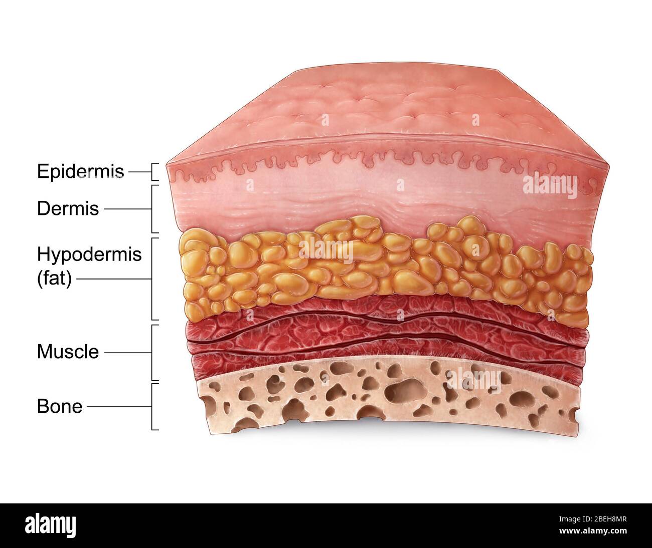
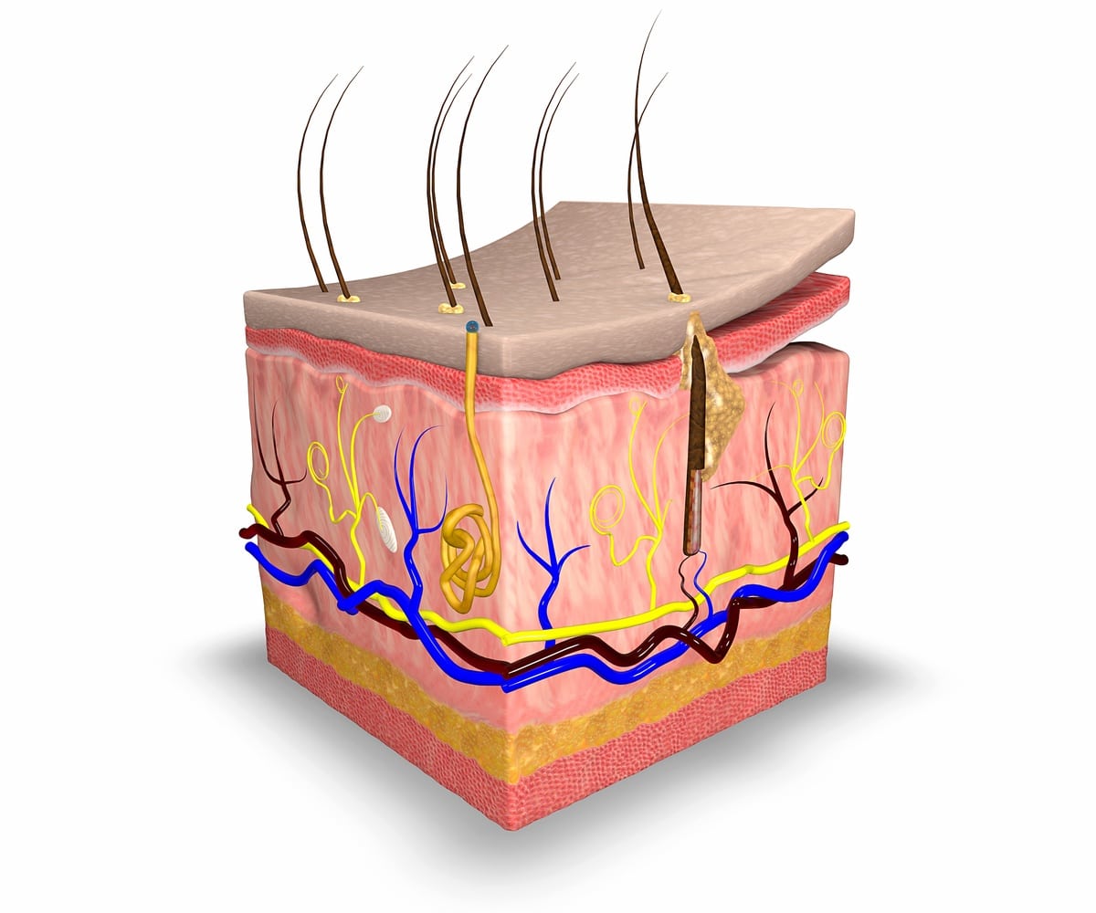
It consists of multiple layers and specialized structures that support its various roles. Below is a detailed breakdown of the anatomy of the skin: Layers of the Skin. The skin is composed of three main layers: the epidermis, the dermis, and the subcutaneous tissue (also known as the hypodermis). Each layer has a distinct structure and specific
Learn about the structure and functions of the epidermis, dermis, and hypodermis, the three layers of the skin. See diagrams, micrographs, and animations of the skin cells and tissues.

Diagram, Structure, Function Biology Diagrams
Learn about the three layers of the skin: epidermis, dermis, and subcutaneous fat layer. Find out how each layer functions and what cells and structures they contain. The skin is the largest organ in the body, covering its entire external surface. The skin has 3 layers—the epidermis, dermis, and hypodermis, which have different anatomical structures and functions (see Image. Cross Section, Layers of the Skin). The skin's structure comprises an intricate network that serves as the body's initial barrier against pathogens, ultraviolet (UV) light, chemicals Skin consists of many layers, made of water, protein, fats and minerals. Skin is the largest organ in the body, protecting it from external elements. Skin consists of many layers, made of water, protein, fats and minerals. Anatomy. What are the layers of the skin? Three layers of tissue make up the skin: Epidermis, the top layer.

Learn about the three main layers of skin (epidermis, dermis, and hypodermis) and their functions. Find out how skin conditions affect the different layers and what to do about them.