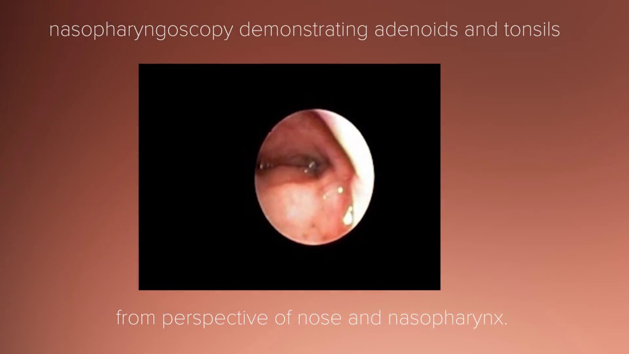
Tonsil and Adenoid Anatomy Review Biology Diagrams Pharyngeal Tonsil. The pharyngeal tonsil refers to a collection of lymphoid tissue within the mucosa of the roof of the nasopharynx.When enlarged, the pharyngeal tonsil is also known as the adenoids.. It is located in the midline of the nasopharynx, and forms the superior aspect of Waldeyer's ring.. The epithelial covering of the pharyngeal tonsil is ciliated pseudostratified epithelium. Pharyngeal tonsil (medial view) The adenoid is a pyramidal shaped structure composed of lymphoid tissue. The apex of this pyramid is extended towards the to nasal septum, and the base sits at the posterior most wall of the nasopharynx. The adenoidal surface is invaginated by a number of folds with some crypts.There is a midline pharyngeal bursa (bursa of Luschka) which extends posteriorly and Enlarged (hypertrophic) tonsils. Larger-than-normal tonsils can block your airway, leading to snoring or sleep apnea. Tonsil cancer. The most common form of oropharyngeal cancer, tonsil cancer is often linked to the human papillomavirus. Symptoms include tonsil pain, a lump in your neck and blood in your saliva (spit).
The two main types of tonsils are the palatine tonsils, which are the ones commonly referred to as "tonsils," and the pharyngeal tonsils, also known as the adenoids. Palatine Tonsils. The palatine tonsils are clusters of lymphatic tissue consisting of lymphocytes, macrophages, and other immune cells.

Tonsils: Anatomy, Definition & Function Biology Diagrams
Objective: This review aims to discuss the basic anatomy and physiology of the palatine and pharyngeal tonsils, with reference to how this foundational understanding may affect patient management and surgical procedures in these regions of the upper airway. Methods: A literature search was performed using PubMed and Google Scholar using the MeSH terms tonsils, adenoids, anatomy, physiology
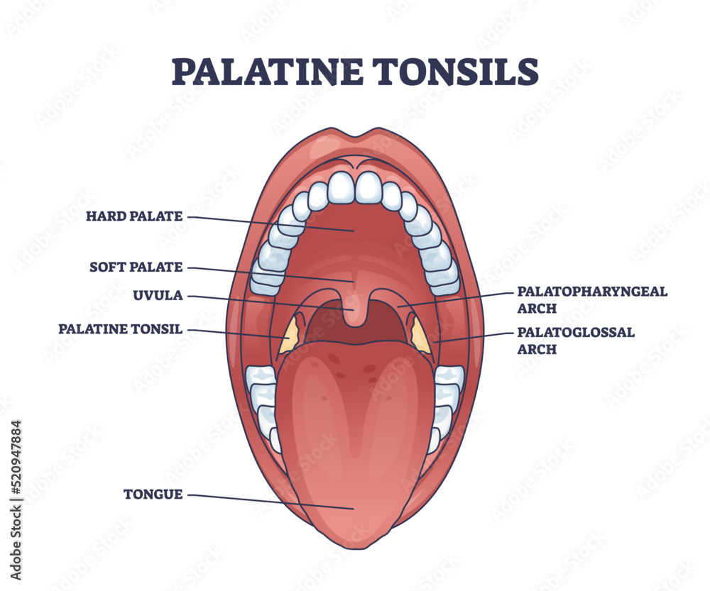
The palatine tonsils are dense compact bodies of lymphoid tissue that are located in the lateral wall of the oropharynx, bounded by the palatoglossus muscle anteriorly and the palatopharyngeus and superior constrictor muscles posteriorly and laterally. The adenoid is a median mass of mucosa-associated lymphoid tissue.

The Tonsils (Waldeyer's Ring) Biology Diagrams
Keywords: Anatomy of tonsils, Adenoids, Waldeyer's ring. The palatine tonsils, adenoids, tubal tonsils, and lingual tonsils are lymphoepithelial tissues that make up the components of Waldeyer's ring, named after the German anatomist Heinrich Wilhelm Gottfried von Waldeyer-Hartz. These entities are together a part of the mucosal immune system. The pharyngeal tonsil, also known as the adenoids, is the most superior component of the pharyngeal lymphoid ring and lies in the superior part (vault) of the nasopharynx.It is attached to the periosteum of the sphenoid bone by connective tissue.The pharyngeal tonsil is covered with ciliated pseudostratified columnar (i.e. respiratory) epithelium. The covering capsule is thinner compared to